
The retina is a thin, light-sensitive layer covering the back inner surface of the eye. Light entering the eye is refracted by the cornea and lens to focus on the retina. The resulting image on the retina is converted into electrical signals that travel to the brain via the optic nerve.
What is the macula?
The macula is a small part of the retina responsible for central and color vision. It enables activities such as reading, driving, and detailed work. The peripheral retina, outside the macula, plays a role in peripheral and night vision.
What does fundus mean in the eye?
The fundus refers to the bottom part of an organ farthest from its opening. In the eye, the fundus includes all structures at the back of the eye, such as the optic nerve, macula, retina, retinal blood vessels, and vitreous. A fundus examination refers to the inspection of this area. Another name for it is the eye bottom examination. Fundus photography is an imaging method that captures these structures and allows them to be archived.
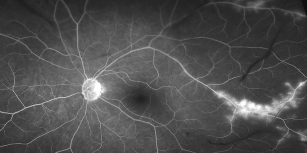
What is fundus fluorescein angiography?
Fluorescein angiography is an eye test used to detect circulation problems in the retina and choroid at the back of the eye. The procedure involves injecting an orange-colored fluorescent dye into a vein and using a special camera to photograph the blood vessels. It is used to diagnose certain eye conditions, including retinopathies and macular degeneration. Fluorescein angiography is a simple and short test that can be performed in a doctor’s office. There is no radiation associated with this test.
Why is fundus fluorescein angiography performed?
This test can detect leakage and abnormalities in the eye’s blood vessels. If there is a circulation problem, it helps outline the affected area for treatment planning. It can also be used to monitor treatment effectiveness.
Fluorescein angiography is used to detect or monitor many conditions, including but not limited to:
• Diabetic Retinopathy
• Macular Degeneration
• Retinal Detachment
• Retinal Vein Occlusion
• Retinitis Pigmentosa
• Retinopathy-Microaneurysms – Abnormal retinal capillaries
• Papilledema – Optic disc swelling
• Hypertensive Retinopathy
• Cancer
• Tumors
• Circulatory Problems
• Inflammation or tumors
How is an eye FFA performed? What is FFA imaging?
For people undergoing FFA:
Your doctor will dilate your pupils using eye drops. You will be asked to place your head on an imaging device and keep it still. A special camera will be used to take photographs. Then, the dye is usually injected into a vein in the forearm, and photographs are taken as the dye flows through the blood vessels in your retina and choroid. Imaging continues for about 10 more minutes.
After the test, your vision may be blurry for a few hours. As the dye leaves your body, your urine will appear bright orange for 24 to 48 hours following the test. Some people may experience mild nausea that disappears quickly due to hypersensitivity to the dye. In some cases, an allergic reaction ranging from mild itching to hives may occur. These reactions usually appear 15 to 20 minutes after injection and are typically treated with observation or antihistamines. Rarely, a more severe reaction may occur requiring urgent medical attention. You may need to stay in the office for about 30 minutes after the test to ensure no reactions occur. Results are available immediately and your doctor can discuss them with you right away.
For more technical details:
FFA requires the use of a special fundus camera equipped with excitation and barrier filters. Fluorescein dye is usually injected intravenously via an antecubital vein at a rate sufficient to produce high-contrast images of the early stages of the angiogram. White light from a flash passes through a blue excitation filter. The blue light (wavelength 465-490 nm) is absorbed by unbound fluorescent molecules, which then emit fluorescent light in the yellow-green spectrum (520-530 nm) at a longer wavelength. A 520-530 nm barrier filter allows only light emitted from the excited fluorescein to be captured. Images are taken immediately after injection and continue for about ten minutes, depending on the pathology observed. Images are recorded digitally or on 35mm film and later evaluated by the ophthalmologist.